X-ray Free-Electron Laser Science
We regularly prepare and carry out experiments at XUV and X-ray Free-Electron Lasers (FELs) around the world, often working within international collaborations. Upcoming projects involve the external page FERMI FEL in Trieste, Italy, the external page SwissFEL in Villigen, Switzerland, external page FLASH and external page European XFEL in Hamburg, Germany, and external page LCLS at SLAC in Stanford, USA.
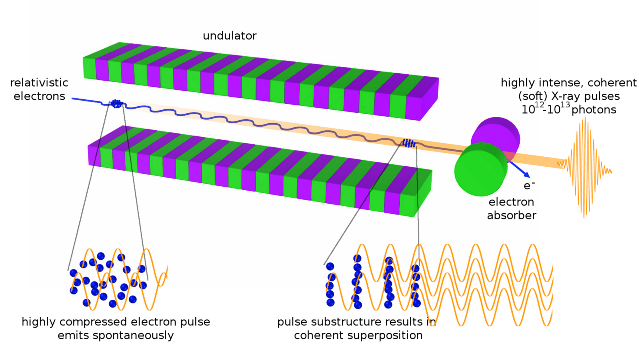
In a simple picture, the intense, coherent, femtosecond pulses from extreme ultraviolet (XUV) and X-ray FELs are produced by accelerating an extremely well compressed bunch of electrons to relativistic velocities and forcing it on a slalom course. The emitted radiation retroacts on the electron bunch, separating it into micro-bunches which then start emitting coherently. XFELs opened up new research fields in physics and far beyond, one of the most prominent examples being coherent diffractive imaging (CDI) of individual, nanoscale specimen such as single viruses or aerosols. In addition, the short pulse durations allow for investigating ultrafast dynamics in a time-resolved fashion.
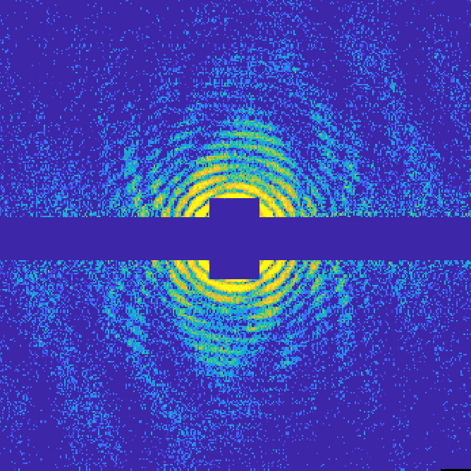
One recent example of an experiment aiming at the determination of nanoscale structures is our 2019 study of Helium-immersed nanostructures at European XFEL. In an international collaboration (with the external page Moeller, external page Vilesov, external page Fennel, external page Meiwes-Broer, and external page Stapelfeldt groups), we imaged helium nano-droplets with tiny clusters and microstructures that grow inside them. The helium droplets are in a superfluid state, therefore the embedded atoms can move without any friction inside the droplets and assemble in special ways, forming nanostructures that cannot be created elsewhere. Coherent diffraction imaging with an XFEL makes the structures and the process of their assemby visible, it's a unique method to study growth under these very extreme conditions.
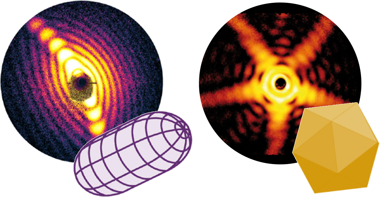
By measuring instead in the long-wavelength end of the X-ray range, the XUV regime, we can trade spatial resolution for three-dimensional information about the shape and orientation of individual nanoparticles. This 3D sensitivity has lead to facinating findings on the shapes of external page silver nanoparticles and external page superfluid helium nanodroplets.
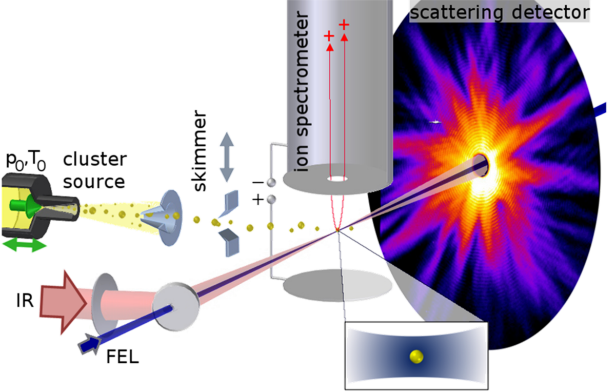
A typical setup of our experiments consists of a combination of single-shot detectors around the focus point of the FEL. A single nanoparticle in free flight is flash-illuminated by a single FEL pulse. The scattered photons form a diffraction image which encodes the nanoparticle’s structure. Other reaction products such as ions, electrons, or fluorescence photons are also measured for every single laser shot, providing further insight into the laser matter interaction. With pump-probe schemes, fast structural changes like ultrafast melting of a metal nanocrystal can be studied with high spatial and temporal resolution.